Role of Strain Echocardiography in Cardiac Amyloidosis
A Case of Advanced Stage TTR Amyloid Heart Disease
In this blog, we will present a patient case of advanced-stage transthyretin (TTR) amyloid heart disease. Cardiac amyloidosis is characterized by the systemic deposition of insoluble proteins in the heart interstitium and includes two primary variants: amyloid light-chain (AL) amyloidosis and transthyretin (ATTR) amyloidosis, both of which can involve the heart. Despite its prevalence, cardiac amyloidosis remains an underdiagnosed cause of heart failure and cardiac arrhythmias, which can lead to delayed diagnosis and treatment.
The diagnostic process is further complicated by the variety of symptoms, with many patients even remaining asymptomatic until the disease reaches an advanced stage. This unique challenge in clinical practice requires a high level of suspicion for a timely diagnosis of cardiac amyloidosis.
While traditional imaging modalities such as basic echocardiography play a pivotal role in identifying typical features like myocardial thickening and pericardial effusion, a more comprehensive assessment incorporating advanced echocardiography techniques such as speckle tracking analysis is essential for early detection and optimal management of cardiac involvement. This approach is crucial, particularly in the early stages of the disease, where subtle abnormalities may provide valuable diagnostic insights and guide management and therapy.
Let’s explore the features of cardiac amyloidosis on echocardiography and speckle tracking with the following teaching case!
Case Study: A 72-year-old male presents with neuropathy, hypotension, and dyspnea. He has elevated levels of NT pro-BNP; he was diagnosed with transthyretin (TTR) amyloid heart disease.
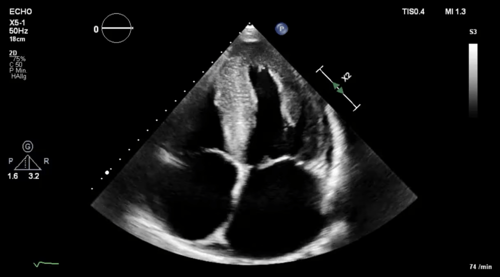
This apical four-chamber view shows a classic (advanced) form of amyloid heart disease, in this case, the patient was diagnosed with TTR amyloid heart disease (the second most common subtype). The echo loop shows the typical features, such as myocardial thickening (symmetric) with a speckled appearance of the myocardium. Note that the left ventricular cavity is small, and the radial function is normal. Still, the longitudinal function, which can be observed by observing the displacement of the mitral annulus, is reduced. The patient also has an enlarged left atrium—atrial enlargement results from impaired left ventricular filling and elevated left atrial pressures. The right atrium is also enlarged. Other findings include a small pericardial fusion. Pericardial effusion is commonly seen in amyloid heart disease, but it rarely has hemodynamic significance. The mitral valve is also somewhat thickened. Finally, it also appears as if the interatrial septum is thickened. The features of amyloid heart disease are so prominent in this patient that the diagnosis of amyloid heart disease is almost certain.
With color Doppler, mitral regurgitation is observed, which is supposedly mild to moderate. It is common to find incompetent mitral, tricuspid, and aortic valves, often thickened due to amyloid deposits in the heart valves. Atrial dilatation also plays a role in the pathomechanism, as it also leads to mitral regurgitation. Although it's rare, patients can develop severe forms of regurgitation.
Along with the typical characteristics of amyloid heart disease ( thickening of the myocardium, mild pericardial effusion, thickened interatrial septum, and left atrial enlargement) and the mild-to-moderate mitral regurgitation, there is also moderate to severe tricuspid regurgitation visible. In amyloid heart disease, tricuspid regurgitation might be caused by amyloid deposits in the valve, postcapillary pulmonary hypertension (left heart disease), dilatation of the right atrial (in particular the annular region), dysfunction of the right ventricle, or a combination of these factors.
Valuable insights with speckle tracking echocardiography
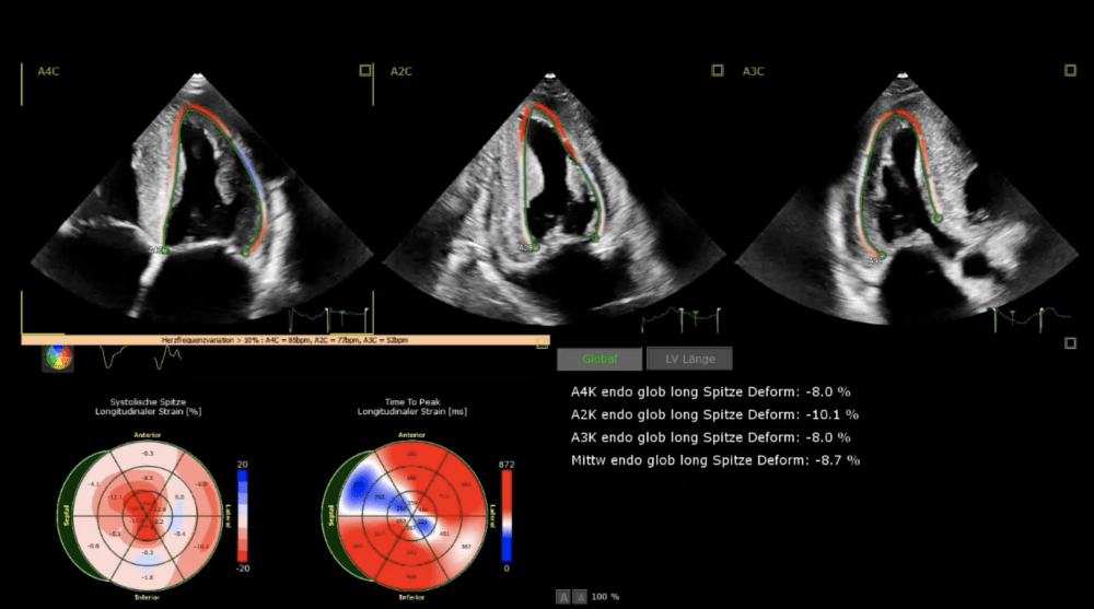
Strain is calculated from a four-chamber view, a two-chamber view, and an apical long-axis view. The bull's-eye display shows the classical characteristics of amyloid heart disease with preserved apical longitudinal strain and reduction of strain in the mid-and basal segments. On the bull's-eye display, we have a typical pattern, which has also been termed cherry on the top (apical sparing). Be aware that apical sparing is not unique to amyloid heart disease but can also occur in other diseases (left frontal hypertrophy). Also, note that global launch to strain is significantly reduced in this example.
From Left to Right: Investigating Right Ventricular Dysfunction in Cardiac Amyloidosis
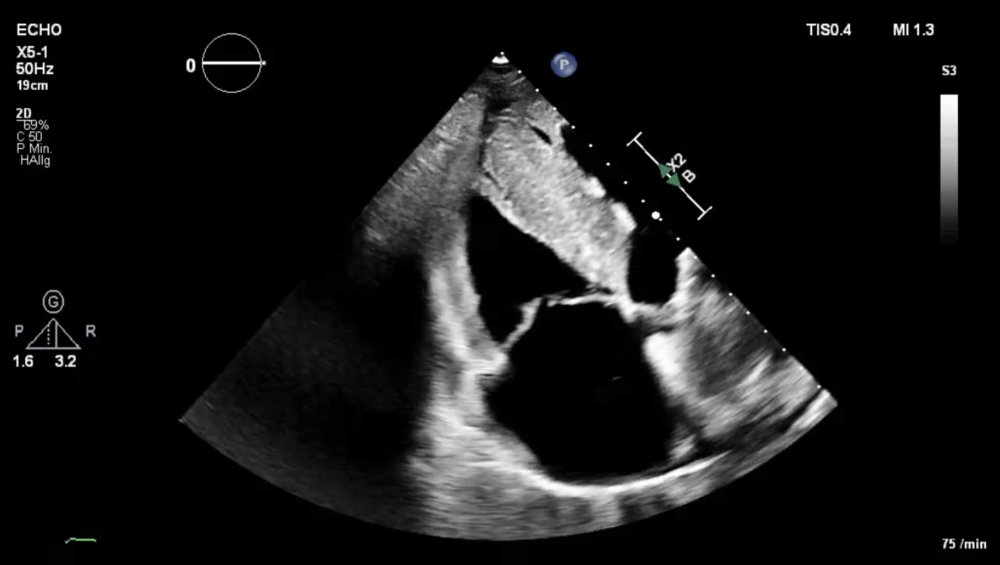
Note that right ventricular function is also moderately reduced, that the wall of the right ventricle is thickened, and that the right atrium is enlarged. Such findings can frequently be found in amyloid heart disease, which affects not only the left but also the right heart.
A right ventricular strain analysis was also performed. The right ventricular free wall strain and right ventricular four-chamber strain (including the septum) are calculated. Both strain values are reduced. This finding indicates reduced longitudinal function of the right ventricle (impaired right ventricular function). Right ventricular strain is commonly reduced in amyloid heart disease.
Additionally, a left atrial strain analysis was done. The algorithm tracks the movement of the left atrial wall and calculates the reservoir strain, conduit strain, and atrial active pump or atrial booster strain. Reservoir strain is a significant measure of atrial function, which is found to be reduced in this patient. This condition is common in amyloid heart disease, where atrial filling pressures are elevated, and amyloid deposits can be found in the left atrial wall. These deposits can appear as focal, multifocal, or diffuse interstitial nodules surrounding cardiomyocytes and replacing normal tissue.
Want to learn more?
Learn how to perform this important modality of echocardiography in our Speckle Tracking MasterClass. You will gain a deep knowledge of strain rate imaging and where it can be applied so that you can use it in your clinical practice immediately. Right now, get an incredible 40% discount on any access times (6, 12, or 24 months) – that is savings up to 854€! Only available until March 17th, so be quick!
Get 40% now


