February is Gallbladder Cancer and Bile Duct Cancer Awareness Month – Listen up!
Gallbladder cancer (GBC) ranks as the fifth most prevalent gastrointestinal malignancy globally and is the most common malignancy of the biliary tract. Sonography is often the initial imaging modality in suspected cases of gallbladder disease due to its cost-effectiveness, widespread availability, and lack of ionizing radiation. GBC arises from the epithelium of the gallbladder, with adenocarcinomas constituting the vast majority (90%) of cases. A crucial risk factor is cholelithiasis, with less than 1% of patients with gallstones developing this form of cancer. Gallbladder polyps with a diameter of ≥ 10 mm pose a risk of malignant transformation. Early-stage diseases are often incidentally discovered during routine cholecystectomy.
The clinical and imaging appearance of gallbladder cancer often overlaps with benign diseases, which makes diagnosis more difficult. In advanced stages, combinations of symptoms such as jaundice, pain and weight loss point more clearly to a malignant cause. Here are some ultrasound findings associated with gallbladder carcinoma (GBC) that should prompt you to examine the patient in detail!
- Gallbladder Wall Thickening: The most frequent ultrasound finding in gallbladder cancer is gallbladder wall thickening, observed in approximately 50% of cases. This thickening may be localized or irregular and is often associated with the presence of gallbladder stones. However, diffuse or focal wall thickening is non-specific and can occur in various gallbladder diseases and extravesicular diseases (such as hepatitis, cirrhosis and pancreatitis). However, certain features such as wall thickening over 12 mm, irregularity, marked wall asymmetry, loss of interface between the gallbladder wall and the liver, and wall calcifications, lymphadenopathy, and bile duct obstructions should raise suspicion for malignancy.
- Gallstones: Gallstones are commonly found in up to 90% of GBC cases. There is a real causal connection, as the presence of gallstones is associated with an increased risk of gallbladder carcinoma due to chronic inflammation/ chronic cholecystis, mechanical damage to the gallbladder wall, conditions such as porcelain gallbladder, and biliary stasis.
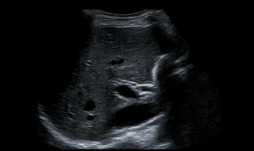
- Biliary dilatation: Gallbladder and bile duct tumors can cause extrahepatic biliary obstruction. So in cases of non-explained extrahepatic biliary obstruction, a malignant cause must be suspected and further assessed with further imaging and lab studies.
- Intraluminal Mass: Not infrequently, gallbladder carcinoma presents as a focal intraluminal mass focal, possibly with wall involvement, with the tumor showing irregular and sometimes ill-defined margins. It can show heterogeneous echotexture and has predominantly low echogenicity.
- Mass Replacing the Gallbladder: Unfortunately, the most common presentation of gallbladder carcinomas is that of a mass replacing the entire gallbladder. It is visualized as a heterogeneous mass with irregular edges, and areas of necrosis or calcification, and may contain echogenic focal points and acoustic shadowing associated with coexisting cholecystolithiasis or wall calcification (think of the so-called porcelain gallbladder!). Although the diagnosis is easy to establish, the clinical options at this advanced stage are limited. There is usually some degree of invasion into neighboring structures or metastatic disease.
How to spot the bile ducts and exclude biliary obstruction during your stand abdominal ultrasound exam?
Unfortunately, due to the mostly asymptomatic nature of these tumors, the presentation is typically late, with the majority of tumors being large, unresectable, with direct extension into adjacent structures or distant metastases present at diagnosis!
While chances look grim, you might be able to initiate the diagnosis earlier through abdominal ultrasound. That is why, for this Awareness Month, we don’t only want to remind you of a thorough look at the gallbladder and the bile duct during your ultrasound exam, but also give you the opportunity to learn how to do it. Our Abdominal Ultrasound BachelorClass can teach you just this – and until February 25th, you can get Lifetime Access for the 6-month standard price!
GET LIFETIME ACCESS NOW
